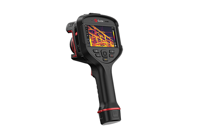
Medical infrared thermography technology is a combination of medical science, infrared thermal imaging technology, and computer multimedia techniques.
How Does Medical Infrared Thermography Work?
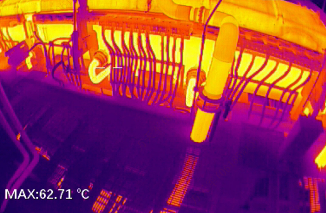
The human body, as a natural heat-emitting organism, has varying temperatures across different regions due to differences in anatomical structure, tissue metabolism, blood circulation, and nervous system activity, creating distinct thermal fields. The IR thermography camera captures far-infrared radiation emitted by the body using an optical-electronic system, filters and focuses it, modulates it, and converts it into electrical signals. These signals are then converted into data via A/D conversion, and after multimedia image processing, the temperature field of the human body is displayed in the form of a pseudo-color thermal image.
Generally, a normal body state exhibits a normal thermal image, while an abnormal state shows an abnormal thermal image. By comparing these differences and combining them with clinical data, it is possible to diagnose the nature and severity of the disease.
What Are the Medical Uses of Infrared Thermography?
1. Combining Structural and Functional Imaging
CT and MRI provide insights into changes in tissue structure, while infrared thermography reveals temperature variations in functional states, such as local blood circulation and neural activity. The combination of structural and functional imaging offers more comprehensive imaging evidence for clinical diagnosis.
2. Identifying Location, Extent, and Severity of Acute and Chronic Inflammation
Inflammation is a common pathological phenomenon, often characterized by redness, swelling, heat, and pain. In clinical practice, while tests like blood work can confirm the presence of inflammation, determining its exact location can be challenging. Infrared thermography helps to easily pinpoint the inflammation site. Acute inflammation presents as areas of high temperature, while chronic inflammation, due to tissue scarring and reduced local blood circulation, typically shows lower temperatures. In cases of chronic inflammation with acute flare-ups, there can be alternating areas of high and low temperatures, providing valuable diagnostic information.
3. Monitoring Blood Flow in Limb Vessels
Infrared thermography is particularly effective for monitoring vascular conditions, especially the blood supply and functional state of limb vessels. In arterial diseases, areas distal to the affected artery will show lower temperatures due to reduced blood flow. In contrast, venous diseases will show higher temperatures in distal areas due to congestion or blood pooling. When a vessel is severed, the region it supplies will exhibit low temperatures, and once blood flow is restored, a rewarming effect will be visible. Compared to other methods such as Doppler ultrasound or skin temperature measurements, infrared thermography is more convenient and intuitive.
4. Early Detection, Monitoring, and Treatment Evaluation of Tumor
Infrared thermography has significant advantages for early tumor detection, continuous monitoring, and treatment assessment. In the early stages of malignancy, when normal cells begin to transform into cancerous ones, they undergo rapid proliferation, accompanied by increased blood circulation to support cell growth. This, combined with the effects of tumor toxins causing local vasodilation, leads to elevated local temperatures. However, in the later stages of a tumor, central necrosis can result in lower temperatures.
Medical infrared thermography, with its high sensitivity, can detect temperature variations as small as 0.05℃, allowing for early identification of abnormal high-temperature areas.


.svg)


_fuben.jpg)

