Did you know that the temperature distribution of a healthy human body exhibits a certain stability and distinct pattern? Different parts of the body have varying temperatures, forming distinct thermal fields. Therefore, when the body becomes diseased, the temperature in the affected area can also change, showing signs of being higher, lower, or irregular. Pain is a common symptom of various diseases, and when pain occurs, it often causes subtle "temperature differences." By detecting these temperature variations, it is possible to identify the location, intensity, and nature of the pain. As a common method of diagnostic technology, infrared thermography can visualize abnormal hot or cold areas in the body. By displaying the health status and potential disease locations through the body's "temperature differences," it makes pain visible and helps doctors pinpoint its cause.
When the Body is Unwell, Temperature Also Changes
As we know, objects in the natural world above absolute zero (-273.15°C) emit infrared radiation, and the human body also emits and absorbs infrared rays. The temperature distribution in a healthy human body is generally stable and characteristic, with different body parts exhibiting different temperatures, creating distinct thermal fields. When a disease or functional change occurs in a part of the body, the blood flow in that area changes accordingly, causing the local temperature to fluctuate. In other words, when a disease develops in a specific part of the body, the temperature in the affected area changes, appearing either too high, too low, or irregular.
Infrared Thermography Camera: Capturing Body Heat for Diagnosis
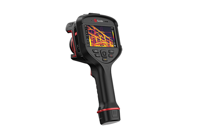
Infrared thermography uses high-precision thermal imaging cameras to capture and reflect these physiological characteristics of the human body. By recording and analyzing the infrared radiation emitted by the body, it presents temperature differences across various parts of the body in visual images. The device is extremely sensitive, capturing the heat radiation produced by cellular metabolism and displaying the abnormal heat sources in the body using different colors. It reveals the distribution, depth, intensity, shape, and progression of these heat sources, helping to locate diseased areas, identify the nature of the illness, and assess the severity of the condition.
Body Temperature Weather Map: Visualizing the Location, Intensity, and Nature of Pain
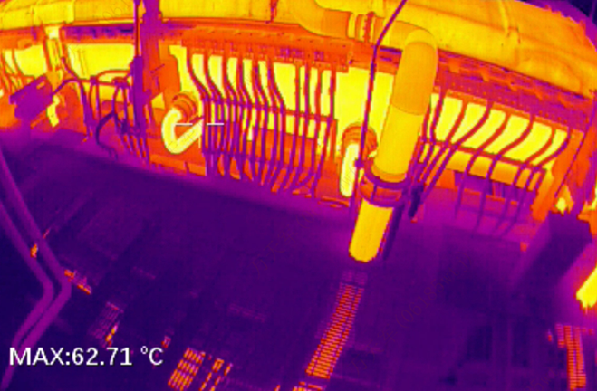
Infrared thermography captures a "temperature weather map" of the human body using an infrared camera. High temperature areas of red color indicate elevated temperatures, while blue areas indicate lower temperatures. High and low-temperature areas in thermographic images are crucial for diagnosing pain-related conditions.
Just a Quick Snapshot: Safe for Pregnant Women and Children
As a diagnostic imaging tool, infrared thermography offers advantages over CT, MRI, and ultrasound, as it is a non-invasive, painless, and non-contact examination. It is also safe and fast. The infrared thermography camera passively receives the infrared radiation emitted by the body, making it harmless with no adverse effects. This makes it safe for pregnant women, children, and the elderly. Furthermore, the imaging process only takes a few seconds, providing a comprehensive scan of the body in a very short time. Infrared thermography not only provides intuitive and reliable evidence for pain diagnosis but also offers valuable clues for early screening of major diseases and health checkups for people with suboptimal health status.
Guide's medical thermography camera allows patients to undergo safe and easy examinations, giving them peace of mind.


.svg)


_fuben.jpg)

