How Does Medical Infrared Thermography Work?
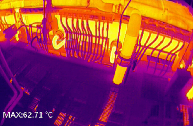
The human body naturally emits heat in the form of infrared radiation. Thermography devices can capture and visualize this radiation, which detects surface temperature and converts it into an image. This technology is based on the principle that changes in skin temperature are closely related to blood flow. When pressure is applied to the skin, even in a healthy individual, blood flow slows in the compressed area, causing the temperature to drop. This is the fundamental working mechanism of medical thermal imaging.
Medical Uses of Infrared Thermography
Medical Infrared Thermography (MIT) is a completely non-invasive diagnostic technique that detects temperature differences on the body's surface. It helps visualize internal diseases or injuries by identifying temperature variations. MIT can be used for early diagnosis, prognosis, and monitoring of treatment progress, allowing for early detection of diseases and prevention of worsening conditions. It is widely used in health screenings, tumor detection, and assessing blood flow to the heart and limbs, as well as in diagnosing pain-related conditions.
With advanced MIT technology, a comprehensive and objective full-body scan can be completed within a few minutes, helping to address potential health issues early, prevent worsening conditions, and tailor treatments to individual needs.
Clinical Analysis of MIT in Temperature Abnormalities
· Increased Temperature (MIT Heat Elevation)
This typically occurs in areas with increased blood flow, such as in cases of acute or chronic inflammation, hyperactivity of tissues, myofascial inflammation, and benign or malignant tumors. However, certain factors such as skin surface irregularities (wrinkles, scars, rashes), recent treatments (physiotherapy, massage, acupuncture), and external factors like mobile phones or backpacks, must be excluded.
· Decreased Temperature (MIT Heat Reduction)
This occurs in regions with reduced blood flow and serves as a warning of potentially dangerous conditions, such as nerve compression or blood vessel blockage. These include myocardial ischemia, brain ischemia, strokes, cervical spondylotic myelopathy, herniated discs, and other nerve compression syndromes.
By integrating MIT with other structural examinations and clinical tests, doctors can visualize the cause of pain, target treatments more effectively, and objectively evaluate therapeutic outcomes. This helps in developing safe, scientifically sound treatment plans and provides an objective foundation for innovative approaches to pain management.
Guide Sensmart: Your Best Choice of Medical Thermography Equipment
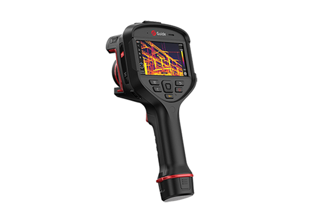
Guide Sensmart's medical thermography equipment boasts its cutting-edge technology and exceptional performance in medical diagnostics. With advanced infrared imaging capabilities, these devices offer a completely non-invasive method to visualize and monitor various health conditions by detecting subtle differences in body surface temperature. This technology enables early detection of diseases, including inflammation, tumors, and circulatory issues, and provides detailed insights into chronic pain, nerve damage, and musculoskeletal disorders.
One of the standout features of Guide Sensmart's equipment is its high-resolution imaging, which ensures precise and detailed thermal images, aiding clinicians in making accurate diagnoses. The devices are also designed with user-friendly interfaces, allowing medical professionals to operate them with ease and efficiency. Additionally, their versatility means they can be used in a variety of medical settings, from general health screenings to more specialized applications like cardiovascular health assessments.


.svg)


_fuben.jpg)

